"Progress in science depends on new techniques, new results, and new ideas - probably in that order." Sydney Brenner
Our group is committed to harnessing and developing new tools in order to enable us to explore new frontiers in experimental neuroscience. We love to combine different techniques - such as two-photon optogenetics and two-photon imaging, patch-clamp recordings and Neuropixels probe recordings - in order to provide complementary information about neural circuit function, and to cross-validate the approaches. We have organized many courses and workshops to disseminate these techniques to the community.
Lab Github site: https://github.com/alloptical
All-Optical INterrogation
Understanding the function of neural circuits requires the ability to not only record the activity of neurons but also to manipulate the activity of the same neurons with the spatial and temporal resolution at which these circuits normally operate. We have developed a new approach for "all-optical interrogation", which combines simultaneous two-photon imaging and two-photon optogenetic manipulation at cellular resolution in the awake cortex. We are using this approach to probe the neural code in cortical and cerebellar circuits.
patch-Clamp REcording
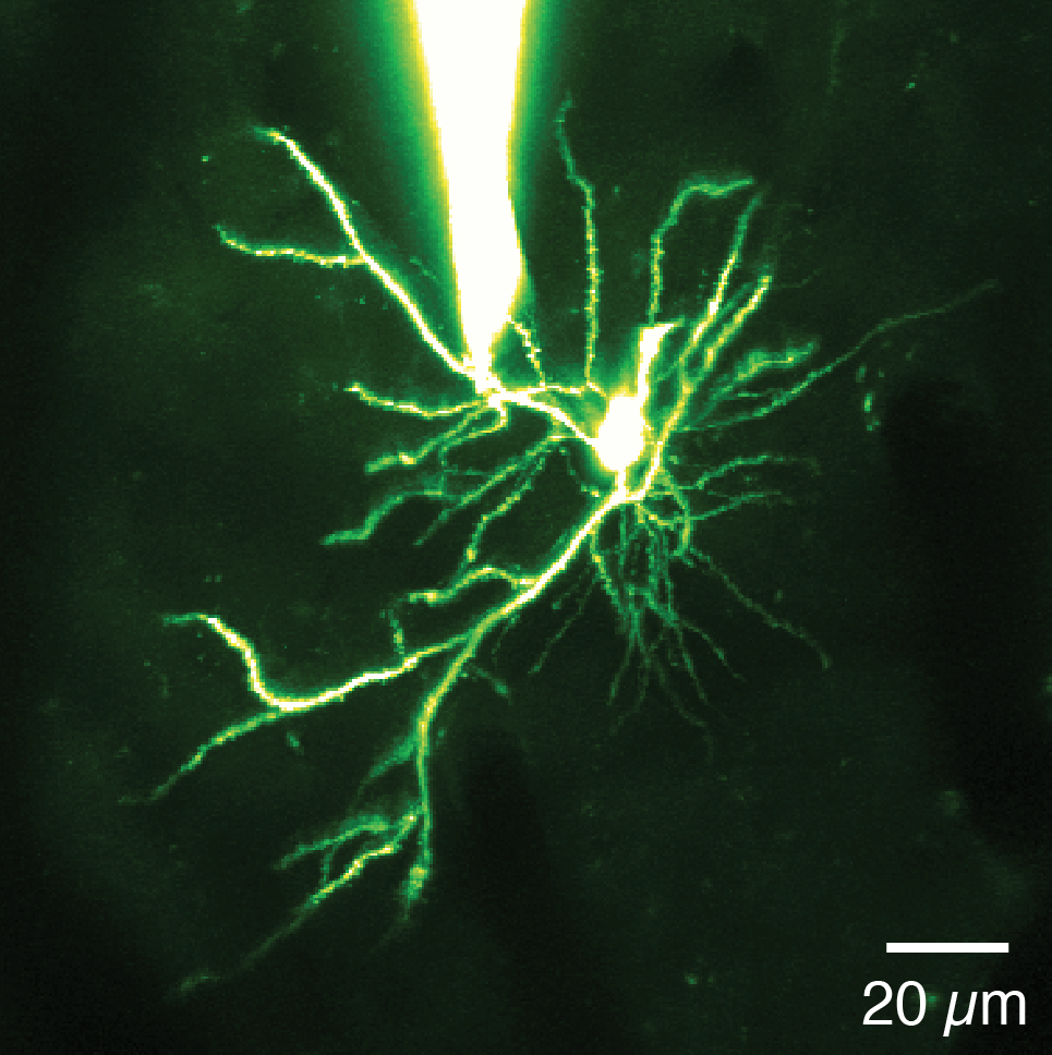
Patch-clamp recording remains the 'gold standard' for electrophysiological investigation of neural activity, providing exquisite sensitivity for recording synaptic input to neurons (using voltage clamp techniques) and for probing subthreshold and supra threshold synaptic integration. We have used patch-clamp recordings to record from axons and dendrites in vivo, and to record from synaptically connected neurons both in vitro and in vivo.
NEUROPIXELS
Neuropixels are next-generation silicon probes with unprecedented capabilities. With 960 densely-spaced sites on a 10 mm long shank, they enable simultaneous recording of the activity of hundreds of neurons across multiple areas of the mouse brain. We are part of a consortium of labs that is testing Neuropixels for use in different brain areas in awake, behaving animals.
HIGH-THROUGHPUT CONNECTOMICS
Understanding a neural circuit requires its wiring diagram - the map of connections between identified neurons (its "connectome'). We are taking advantage of a new approach for visualising connections in neural circuits with ultrastructural resolution using a focused ion beam scanning electron microscope (FIBSEM). This enables us to perform high-throughput imaging of blocks of cerebral cortex or cerebellar cortex, and then trace connections between neurons to define the wiring diagram of these circuits.


